Contents
- 🎵 Origins & History
- ⚙️ How It Works
- 📊 Key Facts & Numbers
- 👥 Key People & Organizations
- 🌍 Cultural Impact & Influence
- ⚡ Current State & Latest Developments
- 🤔 Controversies & Debates
- 🔮 Future Outlook & Predictions
- 💡 Practical Applications
- 📚 Related Topics & Deeper Reading
- Frequently Asked Questions
- References
- Related Topics
Overview
The genesis of radiographic testing is inextricably linked to the discovery of X-rays by Wilhelm Conrad Röntgen in 1895, a breakthrough that earned him the inaugural Nobel Prize in Physics in 1901. Initially explored for medical diagnostics, the potential for industrial applications quickly became apparent. Early pioneers like Michael Pupin and Charles Maggi recognized that these penetrating rays could reveal internal structures of manufactured goods. By the early 20th century, companies like General Electric were developing industrial X-ray equipment. The use of gamma rays, particularly from radium sources, emerged in the 1920s and 1930s, offering a portable alternative for inspecting thicker materials, a development championed by figures like George Herzog and organizations such as the ASTM. This period saw the formalization of RT as a vital tool for quality control in burgeoning industries like shipbuilding and aviation during World War II.
⚙️ How It Works
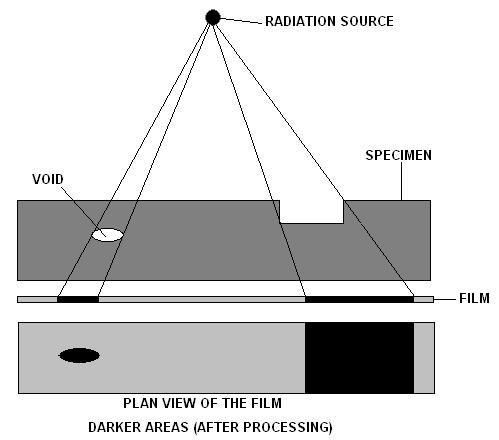
Radiographic testing operates on the principle of differential absorption of radiation as it passes through a material. An X-ray generator or a radioactive isotope (like Iridium-192 or Cobalt-60) emits penetrating radiation. This beam is directed through the test object, and the radiation that emerges is captured by a detector. Denser materials or those with internal flaws absorb more radiation, appearing lighter on the resulting image, while less dense areas or voids allow more radiation to pass through, appearing darker. Detectors can range from traditional silver halide film to intensifying screens, phosphor plates, or advanced digital flat-panel detectors that provide real-time imaging (fluoroscopy) or high-resolution static images. The resulting image, whether film-based or digital, is then interpreted by a certified radiographer to identify and evaluate defects.
📊 Key Facts & Numbers
The global industrial radiography market was valued at approximately $1.5 billion in 2023 and is projected to grow at a compound annual growth rate (CAGR) of around 5.5% through 2030. Over 90% of industrial radiography applications utilize X-rays, with gamma radiography accounting for the remainder, primarily due to its portability. Digital radiography (DR) systems, which offer real-time imaging and immediate data analysis, now represent over 60% of the market share for new equipment sales, a significant shift from film-based systems which dominated prior to 2010. The aerospace industry accounts for nearly 25% of RT usage, driven by stringent safety requirements for critical components. In manufacturing, RT is applied to an estimated 15-20% of critical welds and castings to ensure their integrity.
👥 Key People & Organizations
Key figures in the development of industrial radiography include Wilhelm Conrad Röntgen, whose discovery of X-rays in 1895 laid the foundation. Early proponents of industrial applications included George Herzog, who was instrumental in establishing RT practices in the early 20th century. Organizations like the ASTM (now ASTM International) and the IAEA have been crucial in developing standards and guidelines for RT procedures and safety. Major manufacturers of radiographic equipment include Baker Hughes, Olympus Corporation, and GE Healthcare, each contributing significant technological advancements in X-ray sources and digital imaging systems. The ASNT is a leading professional society that certifies and trains radiographers worldwide.
🌍 Cultural Impact & Influence
Radiographic testing has profoundly shaped industrial quality control, moving it from purely external inspection to internal verification. Its adoption enabled the mass production of reliable components in critical sectors like aviation and nuclear power, directly contributing to enhanced safety records and public trust. The visual language of radiography, with its characteristic grayscale images revealing hidden flaws, has also permeated popular culture, often depicted in fictional scenarios involving medical scans or security screening. The development of portable gamma radiography units, for instance, revolutionized field inspections, allowing for the assessment of pipelines and large structures without the need to transport them to a laboratory. This capability has been vital for infrastructure maintenance and the expansion of global trade through reliable component manufacturing.
⚡ Current State & Latest Developments
The current state of radiographic testing is characterized by a rapid transition towards digital technologies. Real-time digital radiography (DR) and computed radiography (CR) systems are increasingly replacing traditional film, offering faster inspection times, improved image quality, and immediate data archiving. Advancements in portable X-ray generators and miniaturized gamma sources are enhancing field applicability. Furthermore, the integration of artificial intelligence (AI) and machine learning algorithms is beginning to automate defect detection and analysis, promising to increase accuracy and reduce human error. Companies like Vidisco Ltd. are pushing the envelope with ruggedized, portable digital radiography systems designed for harsh environments, while Phoenix X-ray continues to innovate in microfocus X-ray technology for high-resolution imaging of intricate components.
🤔 Controversies & Debates
A significant controversy surrounding radiographic testing involves the inherent risks associated with ionizing radiation. While RT is a powerful diagnostic tool, improper handling or inadequate shielding can lead to occupational health hazards for technicians and potential radiation exposure to the public. This has led to stringent regulatory frameworks, such as those governed by the U.S. Nuclear Regulatory Commission (NRC) and similar international bodies, dictating safety protocols, licensing requirements, and exposure limits. Another debate centers on the interpretation of radiographic images; while digital systems offer enhanced visualization, the subjective element of human interpretation remains, leading to discussions about the reliability and consistency of defect classification and the need for robust training and certification programs like those offered by the ASNT.
🔮 Future Outlook & Predictions
The future of radiographic testing is poised for significant evolution, driven by advancements in digital imaging, automation, and novel radiation sources. We can expect to see widespread adoption of AI-powered defect recognition systems that can analyze images with unprecedented speed and accuracy, potentially reducing the need for highly specialized human interpretation for routine inspections. Miniaturization of X-ray sources and the development of portable, high-energy radiography systems will further expand RT's reach into remote or challenging locations. Emerging technologies like phase-contrast imaging and multi-energy radiography promise to enhance sensitivity for detecting subtle defects. Furthermore, the integration of RT with other NDT methods, such as ultrasonic testing and eddy current testing, within unified digital platforms will offer more comprehensive material characterization. The development of safer, more efficient radioisotopes for gamma radiography also remains an active area of research.
💡 Practical Applications
Radiographic testing finds extensive application across a multitude of industries. In aerospace, it's crucial for inspecting turbine blades, engine components, and airframe structures for fatigue cracks and porosity. The oil and gas industry relies heavily on RT for examining welds in pipelines, pressure vessels, and offshore platforms to prevent catastrophic failures. In manufacturing, it's used to inspect castings, forgings, and welded assemblies for internal defects before they enter the supply chain. The nuclear power industry employs RT to ensure the integrity of reactor components and fuel assemblies. Even in construction, RT can be used to inspect welds in bridges and structural steel. Furthermore, security applications, such as baggage scanning at airports, utilize similar radiographic principles to detect contraband and hazardous materials.
Key Facts
- Year
- 1895 (discovery of X-rays)
- Origin
- Germany (discovery of X-rays)
- Category
- technology
- Type
- technology
Frequently Asked Questions
What is the fundamental principle behind radiographic testing?
Radiographic testing relies on the principle of differential absorption of penetrating radiation. When X-rays or gamma rays pass through an object, denser materials and internal flaws absorb more radiation, while less dense areas allow more to pass through. This variation in radiation intensity is captured by a detector, creating an image that reveals the internal structure and any discontinuities within the material.
What are the main types of radiation used in industrial radiography?
The two primary types of radiation used are X-rays, generated by X-ray machines, and gamma rays, emitted from radioactive isotopes like Iridium-192 and Cobalt-60. X-rays offer controllable energy levels and are typically used for thinner materials, while gamma rays are more portable and better suited for inspecting thicker or denser materials, such as large steel welds.
How has digital radiography changed the field?
Digital radiography (DR) and computed radiography (CR) have revolutionized RT by replacing traditional film. These systems provide immediate image acquisition, enhanced image processing capabilities, and digital archiving, leading to faster inspection cycles and improved defect detection. They also eliminate the need for chemical processing of films and reduce waste, contributing to a more efficient and environmentally friendly workflow.
What are the safety concerns associated with radiographic testing?
The primary safety concern is exposure to ionizing radiation. Technicians must adhere to strict safety protocols, including wearing protective gear, maintaining safe distances, and using shielding to minimize radiation exposure. Regulatory bodies like the U.S. Nuclear Regulatory Commission (NRC) set guidelines for radiation safety, licensing, and monitoring to protect workers and the public.
What kind of defects can radiographic testing detect?
Radiographic testing is highly effective at detecting volumetric defects such as porosity (gas pockets), voids (empty spaces), inclusions (foreign materials trapped within the base material), and cracks. It can also reveal issues with fusion in welds and variations in material density or thickness. The sensitivity to planar defects like cracks can vary depending on the orientation of the crack relative to the radiation beam.
How is a radiographer certified?
Radiographers are typically certified through organizations like the ASNT, which offers various levels of certification (e.g., Level I, II, III) based on training, experience, and passing rigorous examinations. These certifications ensure that individuals possess the necessary knowledge and skills to perform RT safely and effectively, interpret results accurately, and understand relevant codes and standards.
What is the difference between radiography and fluoroscopy?
Radiography typically produces a static, permanent image on film or a digital detector, which is then analyzed. Fluoroscopy, on the other hand, provides a real-time, dynamic view of the radiation passing through the object. This allows for continuous monitoring and immediate observation of changes or movement within the specimen, often used for dynamic processes or live inspection of moving parts.